Occupational therapy and Horner syndrome
 This entry is relatively esoteric, but I spent time researching Horner Syndrome so if the information can help even one more person I suppose that posting it here is worthwhile. This is truly the benefit of the Internet: things that waste space on my hard drive may be a treasure trove of information to someone in the world. Here's hoping.
***
Introduction
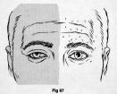
Horner syndrome, also identified as Horner-Bernard syndrome or oculosympathetic paresis, is a constellation of symptoms including miosis, ptosis, apparent enophthalmos, anhidrosis, and reddening of the conjunctiva (Adams &Victor, 1985, p. 208). This condition was described through observations of animals by the French physiologist Claude Bernard. Additionally, some of his co-workers were American surgeons who described this condition in a Civil War soldier who had a cervical sympathetic nerve injury (Kisch, 1951). However, Johann Friedrich Horner, a Swiss ophthalmologist, is generally credited with the first full description of this condition in 1869 despite the earlier publications of the other physicians (Bell, Atweh, Ivy, & Possenti, 2001).
The most notable visual symptoms include miosis, ptosis, and apparent enopthalmos. Miosis is a term for pupillary constriction. In Horner syndrome it is common for one pupil to be more constricted than the other pupil. Under normal conditions, the pupils remain equal at all times in all levels of light. Most references that I scanned indicate that there are no functional implications of anisocoria (having differently sized pupils). However, MacMillan, Cummins, Heron, & Dutton (1994) describe a simultaneous interocular brightness sense test that was sensitive for anisocoria. Additionally, Grossberg & Kelly (1999) describe how binocular brightness sensitivity assists in perception of surface brightness and contour. The functional impact of these issues on everyday vision is not clear, although the same authors have also written about how this process is important for figure ground perception (Kelly & Grossberg, 2000).
Ptosis is drooping of an eyelid. This has an obvious impact on cosmesis but there are also functional considerations. The muscle that is involved by sympathetically-mediated ptosis is the Müller muscle (Adams & Victor, 1985). Ptosis that occurs in the developmental period may have an impact on functional visual acuity as well as visual field (Dray & Leibovitch, 2002).
Enophthalmos is posterior displacement of the eye. In Horner syndrome the narrowing of the interpalpebral fissure, combined with subtle ptosis, causes the appearance of enopthalmos (Lepore, 1983). There are no apparent functional deficits associated with apparent enopthalmos other than the impact on cosmesis.
Etiology
Horner syndrome is caused by an interruption of the oculosympathetic pathway, which is a three neuron pathway. The first order neuron originates in the hypothalamus and descends to the upper thoracic cord where it synapses with second order neurons. These travel over the lung and enter the sympathetic chain in the neck, and synapse in the superior cervical ganglion. Here, third order neurons project postganglionic axons to the eye to innervate the dilator of the iris. Postganglionic sympathetic fibers also innervate the Müller muscle. Postganglionic sympathetic fibers, responsible for facial sweating, follow the external carotid artery to the sweat glands of the face. Interruption at any location along this pathway (preganglionic or postganglionic) will induce an ipsilateral Horner's syndrome (Amonoo-Kuofi, 1999).
Horner syndrome can be caused by damage to first order neurons by conditions including Arnold-Chiari malformation, basal meningitis, basal skull tumors, cerebral vascular accidents, demyelinating diseases, intrapontine hemorrhage, neck trauma, pituitary tumor, and syringomyelia (Bardorf, Van Stavern, & Garcia-Valenzuela, 2001).
Horner syndrome can be caused by damage to second order neurons by conditions including Pancoast tumor, birth trauma with injury to lower brachial plexus, cervical rib, aneurysm/dissection of aorta, subclavian or common carotid artery, central venous catheterization, trauma/surgical injury, chest tubes, lymphadenopathy, mandibular tooth abscess, lesions of the middle ear, and neuroblastomas (Bardorf, Van Stavern, & Garcia-Valenzuela, 2001).
Horner syndrome can be caused by damage to third order neurons by conditions including internal carotid artery dissection, Raeder syndrome, carotid cavernous fistula, cluster/migraine headaches, and herpes zoster (Bardorf, Van Stavern, & Garcia-Valenzuela, 2001).
Additionally, Horner syndrome can be caused by many medications and injuries; the literature is replete with single case studies of esoteric Horner syndrome etiology.
Medical and therapeutic interventions
There are several medical and therapeutic interventions for Horner syndrome. Initially, pharmacologic testing is used to determine the presence of Horner syndrome as well as the possible location of the lesion. The use of 4-10% cocaine eye drops versus 1% Hydroxyamphetamine can help to distinguish between second and third order Horner syndrome. In general, a sympathetically denervated pupil will not dilate to cocaine, regardless of the level of the sympathetic interruption because there is a decreased amount of norepinepherine. Hydroxyamphetamine stimulates the norepinepherine release, so an eye with Horner syndrome with damaged postganglionic fibers (third-order neuron lesions) does not dilate as well as the normal pupil after hydroxyamphetamine drops (Bardorf, Van Stavern, & Garcia-Valenzuela, 2001). Because of the sensitivity of using cocaine in a pediatric population, some authors have advanced the use of 1% apraclonidine for pharmacologic testing, but this does not distinguish pre- or post- ganglionic lesion sites (Bacal & Levy, 2004).
Most of the interventions for Horner syndrome are primarily focused on the cause of the syndrome (disease, injury). However, there are interventions to correct the symptoms of the syndrome. Surgical procedures to correct ptosis have been described in the literature (Glass, Putterman, & Fett, 1990). Interventions for miosis could be made by a referral to an ophthalmologist or optometrist who could fully evaluate the functional impact that the miosis is having on vision.
Several articles in the literature describe physical or occupational therapy intervention for facial paralysis in general, although most of these refer specifically to Bell’s palsy (Beurskens & Heymans, 2003; Cronin & Steenerson, 2003). Some of these methods may be interesting to investigate regarding rehabilitation of muscles of facial expression as they relate to Horner syndrome.
Impact on occupational performance
Horner syndrome may have an impact on occupational performance. Although there is no description of Horner syndrome in the occupational therapy literature, several areas of potential difficulty can be inferred.
One significant area that could be affected includes completion of personal care and grooming occupations because of the cosmetic changes associated with Horner syndrome. Horner syndrome causes unilateral miosis, ptosis, and apparent enopthalmos. These all can physically alter an individual’s appearance and can cause the individual to be self conscious and can cause others to view the individual negatively (Bullock, Warwar, Bienenfeld, Marciniszyn, & Markert, 2001). For this reason alone it is important to address the symptoms of this disorder.
As mentioned previously, visual dysfunction including amblyopia and perhaps visual perceptual problems could develop as a result of uncorrected ptosis and miosis. Visual function has a broad impact on all areas of occupational performance so deficits in these areas could lead to many functional difficulties with participating in preferred occupations.
Another area to potentially consider in the impact of a dysfunctional sympathetic nervous system could have on an individual. Many different conditions cause Horner syndrome, and it is possible that there are other sympathetic nervous system difficulties that are associated with the underlying cause. Occupational therapists are well-trained in understanding sympathetic nervous system functioning and could evaluate this system if needed to determine any impact on occupational performance.
Occupational therapy assessments
Occupational therapy assessments that would be appropriate for an individual who has Horner syndrome include occupation-based assessments that would address issues relating to participation in personal care and the meaning that these occupations have for the individual. The Canadian Occupational Performance Measure (Law, Baptiste, McColl, Opzoomer, Polatajko, & Pollock, 1990) is an individualized outcome measure designed to detect change in a client's self-perception of occupational performance over time. This assessment would be helpful to elicit information from the individual regarding the impact that the Horner syndrome is having on their self-image.
If the individual is a child aged five through nine, The Visual Skills Appraisal (Richards & Openheimer, 1999) could be useful to measure ocular efficiency. There are six subtests that evaluate pursuit, scanning, alignment, locating movements, eye-hand coordination, and fixation unity.
The Motor Free Visual Perception Test (Colarusso & Hammill, 2003) could be a useful test to determine if there are any visual perceptual difficulties. This test measures visual perceptual skills with stimulus items of increasing difficulty or complexity in each of the areas, normed on ages from 4 to adult. The test is used to identify lags in visual processing of spatial relationships, visual closure, and visual memory, which may adversely affect an individual’s functioning.
General observations of the individual completing functional tasks would also be helpful in determining if there are any functional vision deficits associated with the Horner syndrome.
Occupational therapy interventions
Occupational therapy interventions that would be appropriate for an individual who has Horner syndrome include working on self care occupations that could improve the individual’s sense of control over their appearance. This is also an area where it would be interesting to see if any facial exercise programs (Beurskens & Heymans, 2003; Cronin & Steenerson, 2003) would have any impact on the paralysis associated with Horner syndrome.
In addition to the cosmetic impact of ptosis and attempts to strengthen denervated muscles, the individual could learn to compensate for any functional visual field loss by training in scanning techniques (Quintana, 1995, p. 529). These would be particularly helpful for the superior visual field that could be occluded because of eyelid drooping.
If it was determined that the individual had any associated visual perceptual deficits due to loss of figure ground perception, associated perceptual training could be attempted. This training could take the form of remediation or compensatory techniques.
Summary
Horner syndrome is a rare condition that can be caused by a number of medical conditions, many of which themselves can cause visual system dysfunction. Very little is published about Horner syndrome, and most of the available literature discusses esoteric etiology and treatment of the underlying cause of the symptoms. However, the symptoms can not be ignored as they can have a significant impact on a person’s occupational performance.
As very little has been published in the literature on the impact that these symptoms have on function, this makes choosing assessment instruments and interventions a very difficult task. There is still a lot to learn about these symptoms and how they relate to function. There is also a lot to learn regarding the systemic impact of sympathetic denervation, the possible perceptual deficits that can develop associated with uncorrected anisocoria or ptosis, and the role of different intervention techniques to address these deficits. Occupational therapists could help contribute to the body of knowledge regarding this syndrome.
References:
Adams, R.D. & Victor, M. (1985). Principles of neurology, 3rd ed. New York: McGraw-Hill.
Amonoo-Kuofi, H.S. (1999). Horner's syndrome revisited: with an update of the central pathway, Clinical Anatomy. 12, 345-361
Bacal, D.A. & Levy, S.R. (2004). The use of apraclonidine in the diagnosis of Horner syndrome in pediatric patients. Archives of Opthalmology, 122, 276-279.
Bardorf, C.M., Van Stavern, G., & Garcia-Valenzuela, E. (2001, June 26). Horner Syndrome. Retrieved February 15, 2005, from http://www.emedicine.com/oph/topic336.htm
Bell, R.L., Atweh, N., Ivy, M.E., & Possenti, P. (2001). Traumatic and iatrogenic Horner’s syndrome. Case reports and review of the literature. Journal of Trauma, 51, 400-404.
Beurskens, C.H. & Heymans, P.G. (2003). Positive effects of mime therapy on sequelae of facial paralysis: stiffness, lip mobility, and social and physical aspects of facial disability. Otology & Neurotology. 24, 677-681.
Bullock, J.D., Warwar, R.E., Bienenfeld, D.G., Marciniszyn, S.L., & Markert, R.J. (2001). Psychosocial implications of blepharoptosis and dermatochalasis. Transactions of the American Ophthalmological Society, 99, 65-71.
Colarusso, R. P., & Hammill, D.D. (2003). The Motor Free Visual Perception Test (MVPT-3). Navato, CA: Academic Therapy Publications
Cronin, G.W. & Steenerson, R.L. (2003). The effectiveness of neuromuscular facial retraining combined with electromyography in facial paralysis rehabilitation. Otolaryngology - Head & Neck Surgery, 128, 534-538.
Dray, J.P. & Leibovitch, I. (2002). Congenital ptosis and amblyopia: a retrospective study of 130 cases. Journal of Pediatric Ophthalmology and Strabismus. 39, 222-225.
Glatt, H.J., Putterman, A.M., & Fett, D.R. (1990). Müller's Muscle-Conjunctival Resection Procedure in the Treatment of Ptosis in Horner's Syndrome. Ophthalmologic Surgery, 21, 93-96.
Grossberg, S. & Kelly, F.J. (1999) Neural dynamics of binocular brightness perception. Vision Research, 39, 3796-3816.
Kelly, F., & Grossberg, S. (2000). Neural dynamics of 3D surface perception: Figure-ground separation and lightness perception. Perception & Psychophysics, 62, 1596-1618.
Kisch, B. (1951). Horner’s syndrome, an American discovery. Bulletin of the History of Medicine, 25, 284-288.
Law, M., Baptiste, S., McColl, M.A., Opzoomer, A., Polatajko, H. & Pollock, N. (1990). The Canadian Occupational Performance Measure: An outcome measure for occupational therapy. Canadian Journal of Occupational Therapy, 57, 82-87.
Lepore, F.E. (1983). Enopthalmos and Horner’s Syndrome. Archives of Neurology, 40, 460.
MacMillan, E.S., Cummins D., Heron, G. & Dutton, G.N. (1994). The simultaneous interocular brightness sense test. A test of optic nerve function. Archives of Opthamlology, 112, 1190-1197.
Quintana, L.A. (1995). Remediating perceptual impairments. In Trombly, C. (Ed.). Occupational therapy for physical dysfunction, 4th ed. Baltimore: Williams & Wilkins.
Richards, R. & Openheimer, G.S. (1999). Visual Skills Appraisal. Novato, CA: Academic Therapy Publications